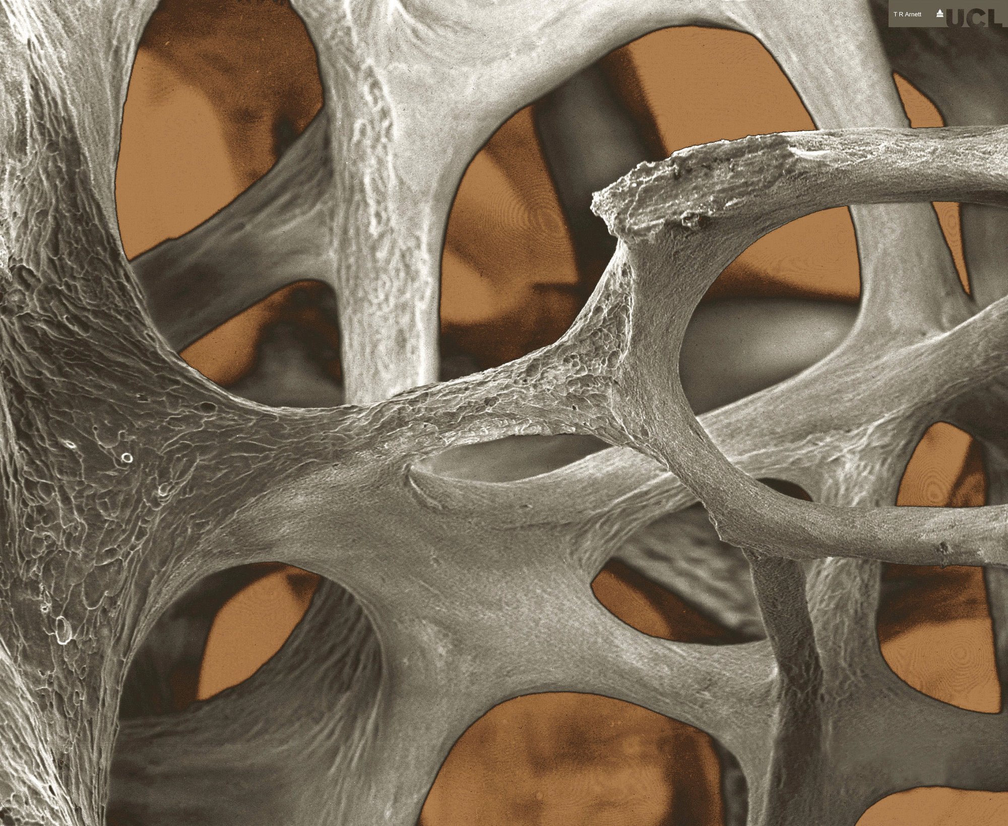
Low-power scanning electron micrograph of osteoporotic bone architecture in the 3rd lumbar vertebra of a 71 yr old woman. Marrow and other cells have been removed removed to reveal eroded bone elements. Field width = 1.4 mm.

By kind permission of Tim Arnett, University College London (t.arnett@ucl.ac.uk)