
By kind permission of Tim Arnett (t.arnett@ucl.ac.uk) & Javier Manzano, UCL.
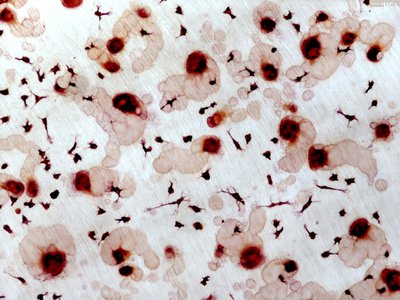
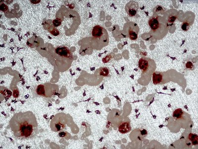
Scanning electron micrograph of activated osteoclast and resorption pit

By kind permission of Tim Arnett (t.arnett@ucl.ac.uk) & Javier Manzano, UCL.
Scanning electron micrograph of activated osteoclast and resorption pit

By kind permission of Tim Arnett (t.arnett@ucl.ac.uk) & Javier Manzano, UCL
Scanning electron micrograph of activated osteoclast and resorption pits.

By kind permission of Tim Arnett (t.arnett@ucl.ac.uk) & Javier Manzano, UCL
Scanning electron micrograph of activated osteoclast and resorption pit.
.
By kind permission of Tim Arnett, University College London (t.arnett@ucl.ac.uk)
.
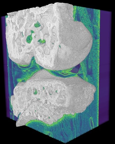
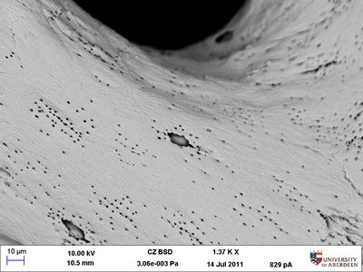
The osteocyte lacunae and canaliculi are also seen within the trabeculae.")
Duncan Bassett, Alan Boyde & Graham Williams
Backscattered electron scanning electron microscope image showing osteoclast resorption of trabecular bone (roughened surfaces).
The osteocyte lacunae and canaliculi are also seen within the trabeculae.
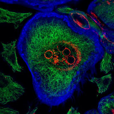
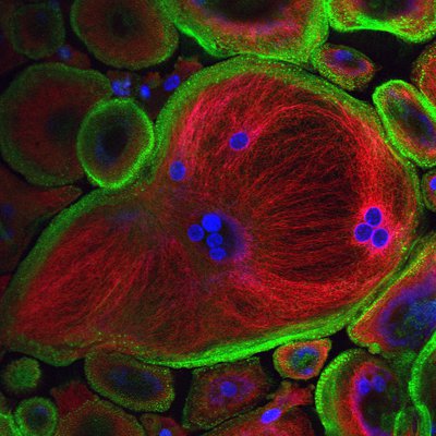
Dr Gudrun Stenbeck, Brunel
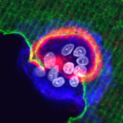
. Cells are stained for osteoclast cell surface marker, integrin alphavbeta3 (green).")
Dr Gudrun Stenbeck, Brunel
Confocal image of human osteoclast culture on bone showing resorption areas enclosed by actin (red). Cells are stained for osteoclast cell surface marker, integrin alphavbeta3 (green).

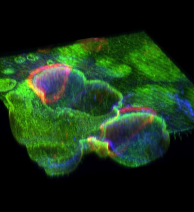
Dr Gudrun Stenbeck, Brunel
3D reconstruction of confocal image of a resorbing osteoclast on dentine revealing the endocytotic organelles of the cell
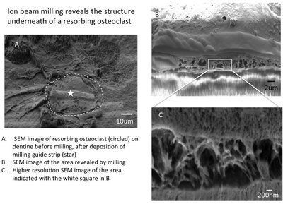
Kevin Mackenzie, Microscopy Facility University of Aberdeen.
Kevin Mackenzie, Microscopy Facility University of Aberdeen.
Dr Fraser Coxon, University of Aberdeen
Dr Fraser Coxon, University of Aberdeen
Dr Fraser Coxon, University of Aberdeen
Dr Fraser Coxon, University of Aberdeen

. The bone is heavily eroded in places by the action of osteoclasts and consists mainly of thin, fragile struts.")
By kind permission of Alan Boyde
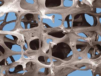
Low power scanning electron microscope image, showing osteoporotic architecture in the fourth lumbar vertebra of an 89 year old woman (x20). The bone is heavily eroded in places by the action of osteoclasts and consists mainly of thin, fragile struts.
. Strong, interconnected plates of bone are visible.")
By kind permission of Alan Boyde
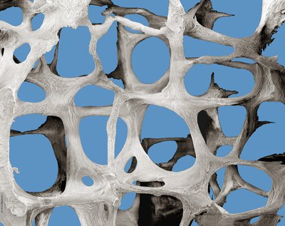
Low power scanning electron microscope images, showing normal bone architecture in the third lumbar vertebra of a 30 year old woman (x20). Strong, interconnected plates of bone are visible.
. A regular pattern of interconnected plates and thick struts of bone can be seen.")
By kind permission of Alan Boyde
Very low power scanning electron microscope image, showing normal bone architecture in the fourth lumbar vertebra of an 41 year year old man (x8). A regular pattern of interconnected plates and thick struts of bone can be seen.

By kind permission of Tim Arnett
Scanning electron micrograph showing osteoclast resorbing bone.

By kind permission of Tim Arnett

By kind permission of Tim Arnett
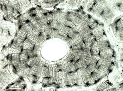
Eruption of adult tooth, cat jaw. H & E stained preparation. Field view 1.5 mm.
By kind permission of Tim Arnett
By kind permission of Tim Arnett

By kind permission of Tim Arnett, University College London (t.arnett@ucl.ac.uk)
Image of normal bone architecture in the 3rd lumbar vertebra of a 30 year old woman.

By kind permission of Tim Arnett, University College London (t.arnett@ucl.ac.uk)
Architecture in the 3rd lumbar vertebra of a 71 year old woman. Note trabecular bone element eroded by osteoclasts.

By kind permission of Tim Arnett, University College London (t.arnett@ucl.ac.uk)
Architecture in the 3rd lumbar vertebra of a 71 year old woman. Note trabecular bone element perforated by osteoclasts.

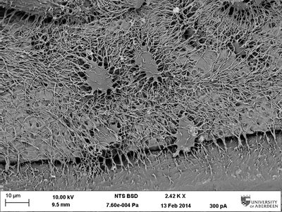
By kind permission of Bram Sengers and Richard Oreffo
Human bone marrow stromal cells spreading on trabecular bone.

By kind permission of Tim Arnett, University College London (t.arnett@ucl.ac.uk)
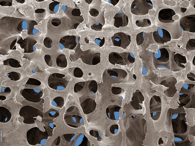
Low-power scanning electron micrograph of osteoporotic bone architecture in the 3rd lumbar vertebra of a 71 yr old woman. Marrow and other cells have been removed removed to reveal eroded bone elements. Field width = 1.4 mm.

By kind permission of Bram Sengers and Richard Oreffo
Human bone marrow stromal cells spreading on trabecular bone.

By kind permission of Tim Arnett.
Low-power image of osteoporitic bone architecture.
 Marrow and other cells have been removed. Extensive pitting caused by osteoclasts in the osteoporotic bone (lower panel)")
© Tim Arnett, University College London (t.arnett@ucl.ac.uk)
(3rd lumbar vertebrae) Marrow and other cells have been removed. Extensive pitting caused by osteoclasts in the osteoporotic bone (lower panel)
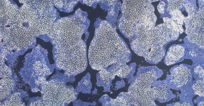
. Trabecular bone structure is particularly well preserved in ribs.")
© Tim Arnett, University College London (t.arnett@ucl.ac.uk)
Section of ichthyosaur vertebrae and ribs in pyrite nodule. Seatown, Dorset, UK (190 myr). Trabecular bone structure is particularly well preserved in ribs.
© Tim Arnett, University College London (t.arnett@ucl.ac.uk)
© Tim Arnett, University College London (t.arnett@ucl.ac.uk)
© Tim Arnett, University College London (t.arnett@ucl.ac.uk)

Please email directly for re-use permission")
Prof Peter Zioupos
© Peter Zioupos, University of Hull (p.zioupos@hull.ac.uk)
Please email directly for re-use permission

Please email directly for alternative formats and re-use permission.")
Prof Peter Zioupos
© Peter Zioupos, University of Hull (p.zioupos@hull.ac.uk)
Please email directly for alternative formats and re-use permission.

Please email directly for alternative formats and re-use permission.")
Prof Peter Zioupos
© Peter Zioupos, University of Hull (p.zioupos@hull.ac.uk)
Please email directly for alternative formats and re-use permission.

Please email directly for alternative formats and re-use permission.")
Prof Peter Zioupos
© Peter Zioupos, University of Hull (p.zioupos@hull.ac.uk)
Please email directly for alternative formats and re-use permission.

Please email directly for alternative formats and re-use permission.")
Prof Peter Zioupos
© Peter Zioupos, University of Hull (p.zioupos@hull.ac.uk)
Please email directly for alternative formats and re-use permission.